Square drops of colored water in a grid pattern. The surface on which each four mm drop was placed is chemically patterned so that drops form squares. Research: G.M. Whitesides, Harvard University. Photo by Felice Frankel.
A few months ago, a particularly breathtaking astronomical image appeared on the front pages of newspapers around the globe. These stunningly colored shapes captivated those who saw them. The picture, captured by the Hubble Space Telescope, was of the Cone Nebula, but the colors in the image were not actually seen by astronomers. They were, in fact, a construction from three separate sets of digital readings taken in blue, near-infrared, and hydrogen-alpha filters.
Interestingly, that Cone Nebula image is fundamentally the same as the one of nanowires that I made with a scanning electron microscope. Both of these pictures are visual displays of numbers; they are representations of data. My nanowire image is a manifestation of the data made with image-processing algorithms that enable us to see structure. After collecting that data, I digitally colored the image. By doing this, I was essentially changing the data, just as the astronomers did when they balanced the values of their three-data sets to create a readable image. But in this case, the color choices I made had nothing to do with the material. Mine were purely aesthetic decisions that provided representations to help create a more engaging image than the original.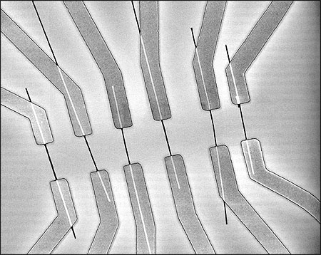
Nanowires, measuring 2-5 nanometers, span across electrodes. Image taken with a scanning electron microscope. Research: C. Lieber, Harvard University.
As scientific research becomes more visual with the advancement of sophisticated image-capture techniques, we are now able to “see” beyond what our unaided eyes allow. Like Hubble, although on a completely different scale, an atomic force microscope or a scanning electron microscope (which I used for the nanowire picture) can capture images and analyze structures never before seen. Now they can be measured in nanometers, 10–9 meters or a billionth of a meter. And with other new instruments we are able to detect the femtosecond, 10-15 of a second, allowing us to comprehend the enormity of this diminutive flash of time and begin to “see” molecules in motion and study how they operate.
That such images can be “constructed” does not diminish their value in increasing our appreciation for and understanding of the scientific advances they reveal. Scientific pictures previously made on film, for example, are as much of a construction and a representation of reality, only made with silver instead of pixels. The adjusted colors of the Cone Nebula are not simply imagined, rather they were created through the human interpretation of technological data. Gaining this kind of extraordinary view offers us a remarkable window, but opening it requires adherence to the scientific integrity of the process. Certainly the misuse of this technology is possible, but so it was with film. It is just easier to manipulate images in digital form. Journalists should simply be more aware of this as images of all kinds come into wider use.
Even as these previously unfathomable concepts are clarified through advancements in imaging and technology, we are still in desperate need of new vocabularies to better communicate these ideas among ourselves, as scientists, and to the public. And in the years ahead, pictures will assume an increasingly prominent role in communicating scientific information. When published along with the drawings and illustrations currently made by graphic artists, images will become powerful tools in making difficult concepts accessible and inspiring to the non-scientist.
These technological tools will result in the emergence of an entirely new community of image-thinking writers and editors, illustrators, information architects, photographers and scientists, along with a fresh approach to science journalism. The creative process of telling scientific stories will become a collaboration among writers, editors and picture makers. Together they will develop a new and rigorous visual vocabulary of science as a compliment to the written word.
This new process in science communication will produce a different kind of journalistic thinking by contributing richer and more informative visual tools not only to the public, but also to the research community as a whole. And with this new thinking will emerge a well deserved, if belated, respect for the power of the image.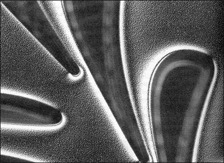
A microscopic image of a detail of a microrotor. The large curved blade measures about fifty microns cross. Research: A. Epstein, MIT. Photos by Felice Frankel.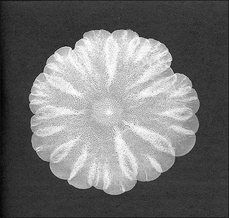
A colony of yeast in a floral arrangement in a petri dish. Research: T. Reynolds, G. Fink, MIT’s Whitehead Institute. Photos by Felice Frankel.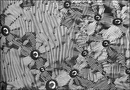
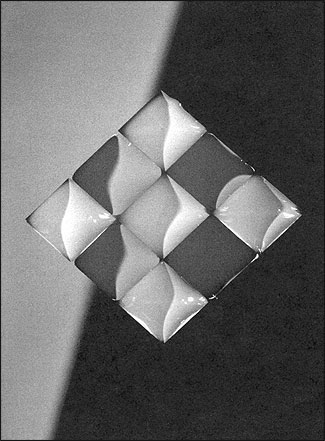
A thin layer of gold buckling in patterns on the surface of plastic. Photo taken under a microscope. Research: N. Bowden, G. M. Whitesides, Harvard University.
Science photographer Felice Frankel is a research scientist at the Massachusetts Institute of Technology. Her recently published book, “Envisioning Science: The Design and Craft of the Science Image” (The MIT Press, 2002), guides researchers and students in how to produce more communicative science images. She is director of the Envisioning Science Project at MIT and is coauthor with George M. Whitesides of “On the Surface of Things: Images of the Extraordinary in Science” (Chronicle Books, 1997). Each of the images reproduced here were originally in color. Her work can be found at web.mit.edu/felicef.